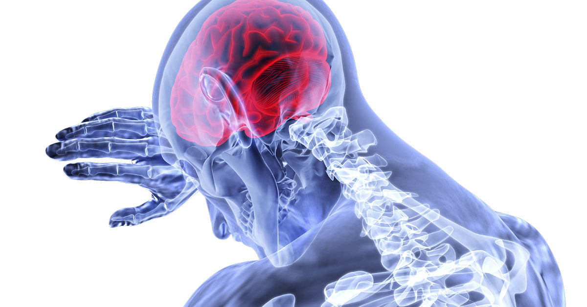
Craniotomy would be needed by patients suffering from conditions within skull, like aneurysms.
Craniotomy for the Treatment of Aneurysms

What is an Aneurysm?
An aneurysm is a weak area in the wall of a blood vessel that causes bulging, dilation or ballooning of the blood vessel. In most cases, aneurysms form at the point where blood vessels divide into branches. A cerebral aneurysm is an aneurysm in a blood vessel of the brain. An aneurysm can exist in a ruptured or unruptured state. A ruptured aneurysm is one that has broken open.
What causes Aneurysms?
Aneurysms in brain vessels occur when an area of the blood vessel's wall is weakened. Aneurysms can be congenital, meaning that they are present from birth, or they may develop later in life. The blood vessel wall might be weakened due to infection or injury. Some people have hereditary disorders that make it more likely that they will have an aneurysm. Some of these disorders include polycystic kidney disease, Osler Weber Rendu, and Marfan's.
Risk factors that may lead to or facilitate the development of aneurysms include high blood pressure, diabetes, genetic disorders, injury or trauma to blood vessels, complication from a blood infection, and smoking or usage of nicotine. Aneurysms are found in 2 to 7 percent of the population.
Symptoms and Diagnosis
The symptoms of aneurysms depend on whether they are ruptured or unruptured.
Patients suffering from a ruptured cerebral aneurysm may exhibit symptoms such as: severe headaches, nausea, stiffness in neck, vision problems, sensitivity to light, called photophobia, loss of sensation, and vomiting. A rupture can cause a person to go into a coma and can even kill the person.
Most patients with unruptured cerebral aneurysms do not show any symptoms. However, some may experience cranial nerve palsy, double vision or visual loss, sudden severe headache, numbness in any part of body, dilated pupils, progressive weakness or neck pain.
The following are used to diagnose aneurysms: cerebral angiography, computed tomographic angiography, or CTA, computed tomography, or CT, scans, doppler ultrasound, and magnetic resonance angiogram, or MRA.
Treatment Options for Aneurysms
Not all aneurysms require immediate treatment. Very small, usually less than 3 mm, aneurysms are less likely to break or rupture, so they may not need immediate treatment. A surgeon may decide to treat aneurysm, even when it does not show any symptoms, in order to prevent the possibility of a fatal rupture in the future.
Aneurysms can be treated either by surgery or by catheter angiography methods. In surgery, the head in opened and the aneurysm is clipped so no blood flows into it. In coiling, a catheter is placed into the artery and titanium coils are placed inside the aneurysm, again preventing blood flow into the aneurysm.
A craniotomy is a surgical procedure that involves removal of a portion of the skull, or cranium, to access the brain. This surgery is performed by a neurosurgeon while the patient is under anesthesia. A craniotomy can be performed on any part of the skull depending on the location of the brain that needs to be accessed. The portion of the skull that is cut out is called a bone flap.
How is a Craniotomy for an Aneurysm Performed?
The following outlines the steps involved in performing a craniotomy:
Before the Procedure
The patient lies on an operating table and is given general anesthesia. Their head is then placed in a device to keep their head in a fixed position during the procedure.
Initial Incision
Some hair may be shaved to clear the incision area. The surgeon makes an incision in the skin. Next, the skin and surrounding muscles are moved aside.
Exposing the Dura
The bone in the exposed part of the skull is cut and the bone flap is lifted. This exposes the covering, called the dura, that protects the brain tissues.
Cutting the Dura
The dura is cut and the blood vessel with the aneurysm is located.
Locating and Treating the Aneurysm
The aneurysm itself is located and treated by placing a titanium clip around the base and occluding it. Then, the surgeon will confirm that blood is still flowing normally in the vessels around the aneurysm.
Ending the Procedure
The dura is closed and the bone flap is replaced. Finally, the skin is restored to its original position and sutured.What Happens After the Craniotomy for an Aneurysm is Performed?
After surgery, the patient is moved to an intensive care unit, or ICU, for close monitoring of their vital signs. They may stay on ventilation and breathing tube for some time. After the procedure, the patients may have a headache or feel nauseated. The patient will remain in the hospital for a few days and receive some short-term rehabilitation.


